Precision Disc Treatment
A herniated disc, where part of the disc protrudes through a tear in its outer layer and compresses nearby nerve structures, can cause various problems. This can lead to pain, numbness, or weakness. Treatment options include physiotherapy, pain management, and, in severe cases, surgery.
Location of the Herniation
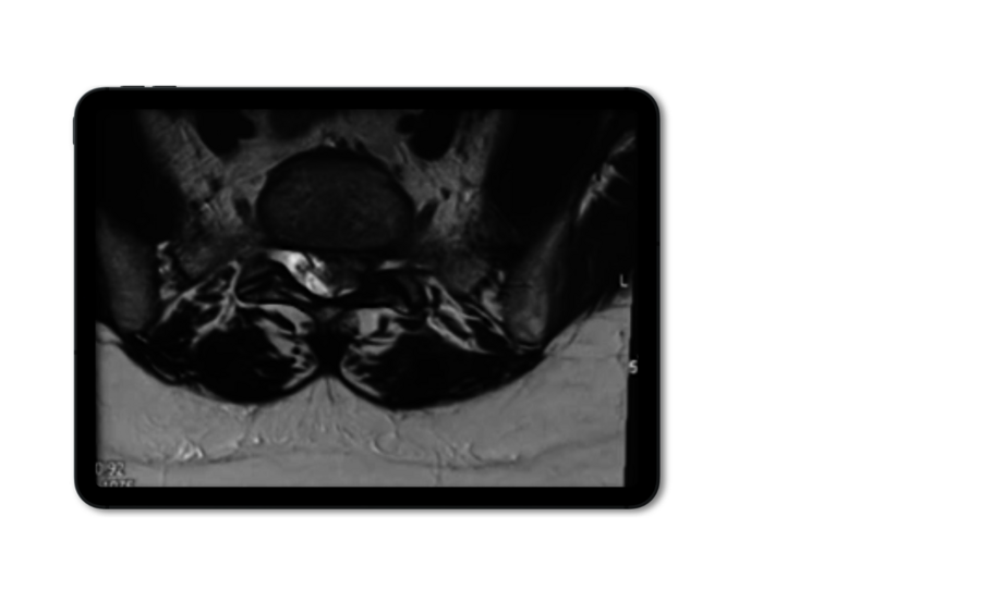
Interlaminar Disc Herniation
Depending on the level of the disc herniation in the spine, the interlaminar technique can be used to treat all types of medial herniations, even migrated herniations.
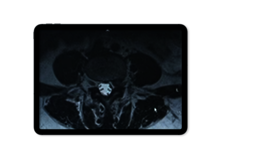
Foraminal Disc Herniation
A foraminal approach is first choice for paramedial and foraminal hernias, unless other criteria such as foraminal stenosis contraindicate.
Techniques
Lumbar
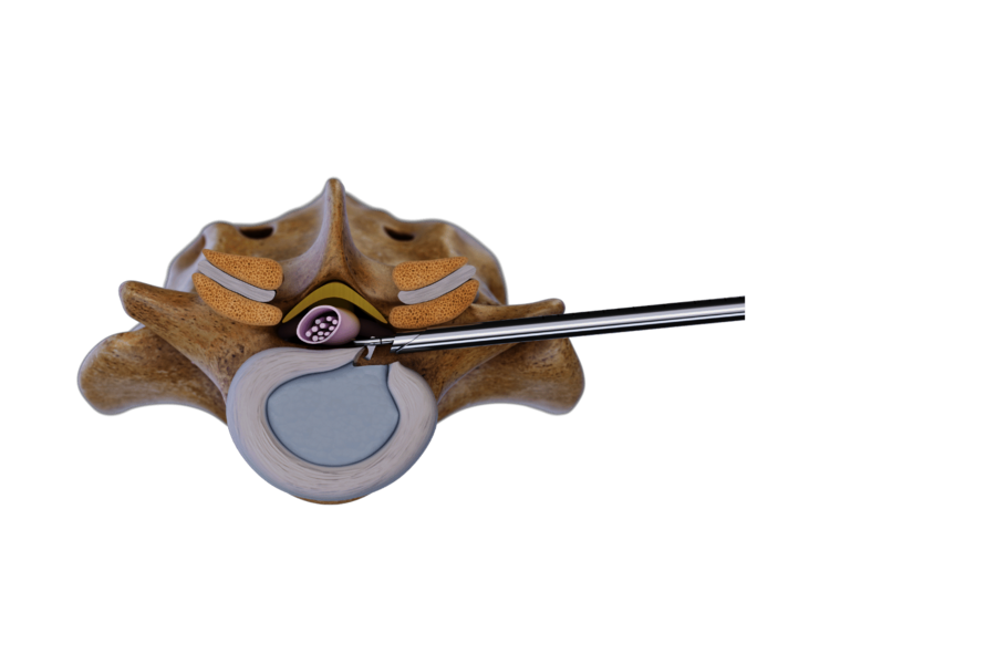
Foraminal technique
Foraminal approaches are minimally invasive techniques for treating spinal pathologies, located extra- and intraforaminally as well as inside the spinal canal and disc. It offers direct decompression with minimal surgical trauma. Depending on pathology, different access angles are used:
- Lateral Intra- and transforaminal approach for treating pathologies, located inside the intervertebral foramen and/ or inside the spinal canal: Angled at ~20°
- Posterolateral transforaminal approach”: Angled at ~45°, ideal for central disc pathologies. Narrow intervertebral foramina may require bone removal using manual or motorized burrs to ensure access.
- Extraforaminal approach for treating extraforaminal disc herniations and foraminal stenosis in lumbar and thoracic spine segments.
Using a lateral-to-posterolateral access angle (20°–30°), the puncture cannula is anchored in the caudal pedicle. The VERTEBRIS diskoscope and instruments are advanced cranially under full-endoscopic visualization to remove the herniated disc safely.

Interlaminar technique
The interlaminar technique accesses the spinal canal through the interlaminar window. This is usually possible at the L5/S1 level without additional bone material removal. In higher lumbar spine levels and in stenosis cases an additional bone resection is required. This technique is ideal for intraspinal herniations and pathologies, particularly in cases of migrated disc herniation or stenosis.
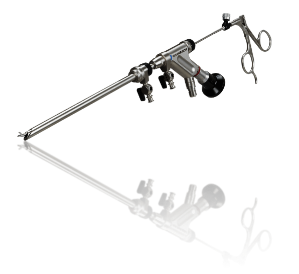
Precision-controlled Surgery
VERTEBRIS Lumbar
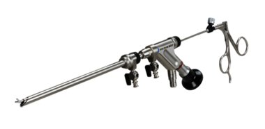
High-resolution endoscopes enable minimally invasive spine surgery in foraminal and interlaminar techniques. The optimized design reduces tissue damage, while advanced fluid management enables clear high-resolution visualization. Specialized instruments ensure precise removal of soft tissue and bone, including by radiofrequency ablation.
Techniques
Cervical
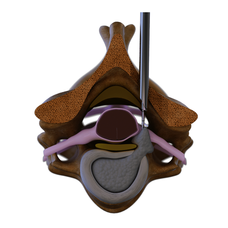
Posterior cervical Surgical Technique
The full-endoscopic posterior approach is mainly for mediolateral to lateral herniated discs. This 7mm diameter access allows atraumatic soft tissue passage to reach the laminae and facet joint. Using high-resolution endoscopic imaging, mechanical burrs remove parts of the laminae and facet joint to locate and remove the herniated disc and relieve the nerve root.

Anterior transdiskal surgical technique
This approach treats medially to mediolaterally localized herniated discs not accessible via the posterior approach. After palpating and passing ventral structures (e.g., esophagus, arteries), a thin dilator is inserted into the disc under X-ray guidance. A special oval dilation system advances to the dorsal edge of the cervical disc. Through an access sleeve, the herniated disc is removed transdiscally under endoscopic visualization.
High-quality Products
See matching systems
VERTEBRIS
lumbar
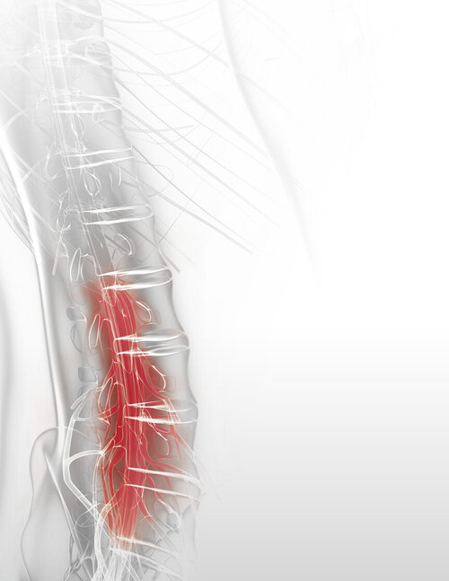
Procedures can be performed using foraminal or interlaminar approaches.
More information

VERTEBRIS
cervical
Two specialized instrument sets, designed for anterior and posterior approaches.
More information
