Advanced Stenosis Solutions
Neural structures within the spine may become compressed by bony or ligamentous tissues within the spinal canal. Categorization based on location of the stenosis is used for differentiating lateral (foramen, lateral recess) and central spinal stenosis.
Location of the Stenosis
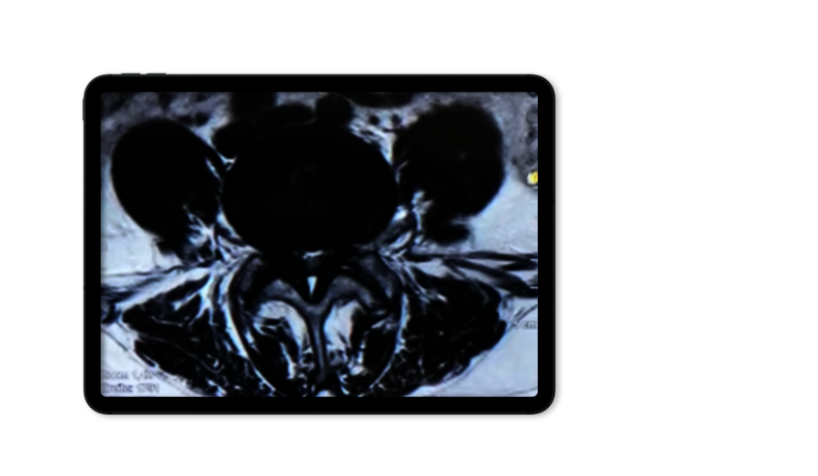
Central Stenosis
The central stenosis involves compression of the spinal cord and nerve roots. Symptoms can be central or radicular. Surgical treatment includes resecting impinging bone and the ligamentum flavum via an ipsilateral full-endoscopic interlaminar approach, extending to the contralateral side when necessary. The VERTEBRIS stenosis system ensures effective bilateral decompression while preserving non-compressive tissues.

Foraminal Stenosis
The foraminal stenosis compresses the exiting nerve root, causing radicular symptoms. It is due to degenerative changes in the facet joints and pedicles. Treatment includes foraminal bone resection using a full-endoscopic foraminal approach (extra- or intraforaminal) with special tools like the Tip-Control Articulating Burr for widening the foramen. Neural structures are identified and protected throughout the procedure.
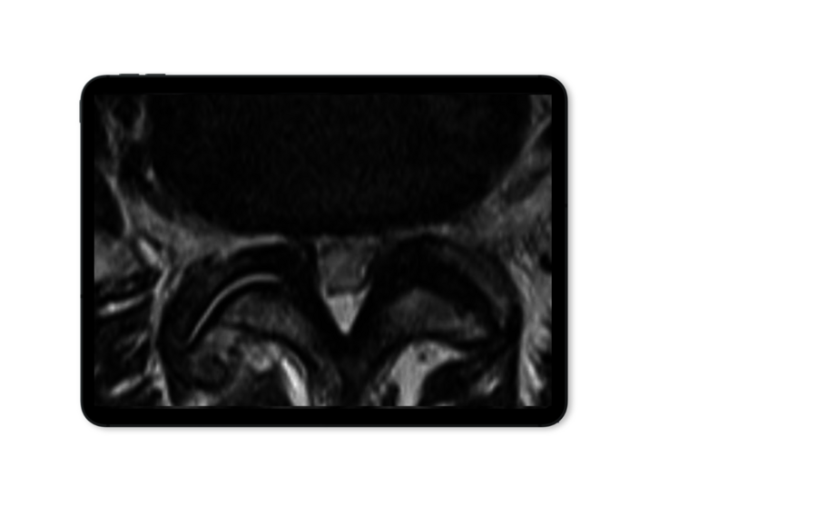
Lateral Recess Stenosis
The lateral recess stenosis causes compression of the traversing nerve root and lateral portions of the spinal cord. This is caused by a narrowing of the bony structures in the lateral recess of the spinal canal.
Surgical intervention involves precise bone resection via a full-endoscopic interlaminar approach using the VERTEBRIS lumbar/ stenosis system. Steps include resecting the descending articular process and carefully opening the ligamentum flavum to expose and decompress the neural structures.
Techniques
Spinal Canal Stenosis
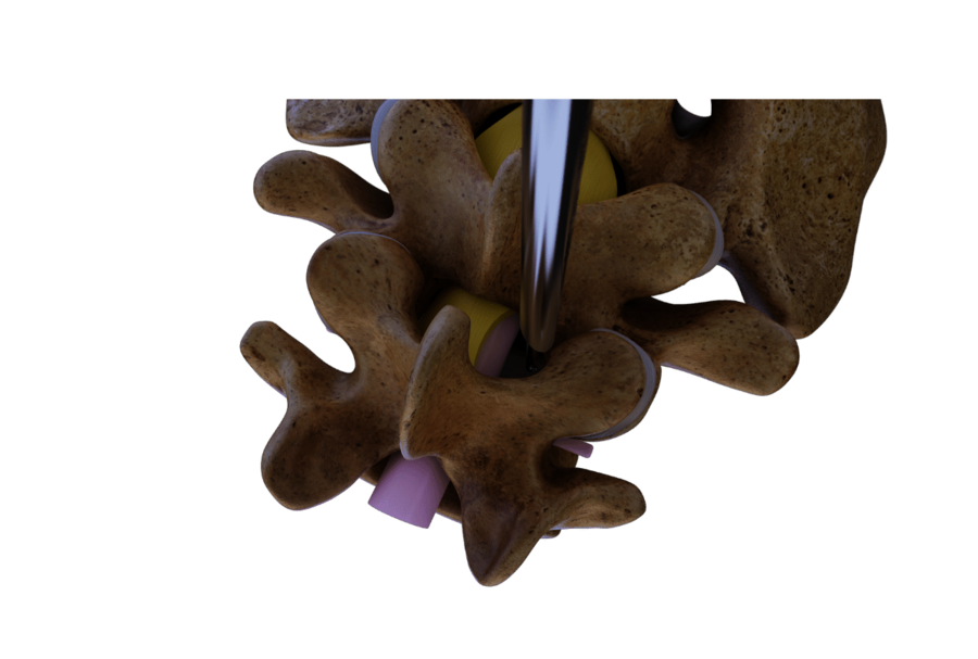
On one side
Ipsilateral decompression
Ipsilateral decompression begins with exposure of the bony structures and continues with resection of the descending facet, lamina, and ligamentum flavum. In the case of central stenosis, the procedure extends to the midline. Neural decompression must be ensured cranially, caudally, and laterally.
In some cases, a ventral epidural annulus or osteophytes may need to be checked and removed if symptomatic.

Over-the-top Technique
Contralateral decompression
Contralateral decompression for bilateral central stenosis involves an "over-the-top" technique. A unilateral approach is employed, starting with ventral resection of the spinous process to access the contralateral side. Laminotomy and partial facetectomy are performed before fully resecting the ligamentum flavum.
Care is taken to avoid excessive retraction of neural structures to minimize neurological damage.
High-quality Products
See Matching Systems
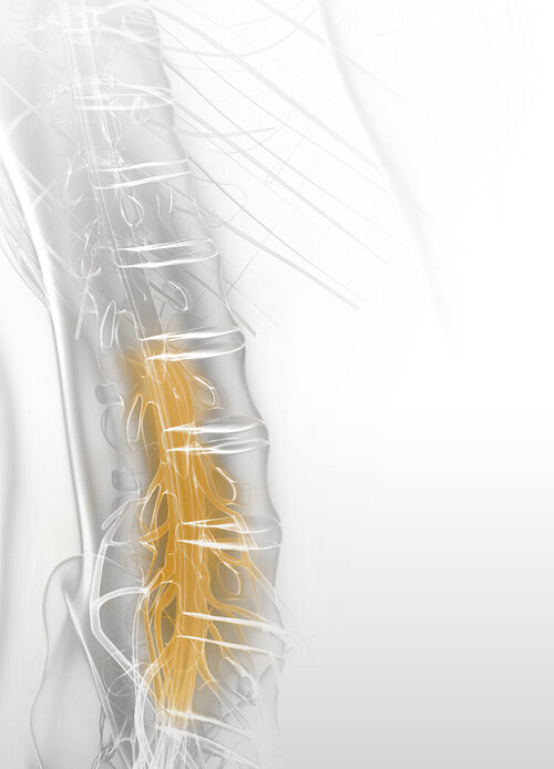
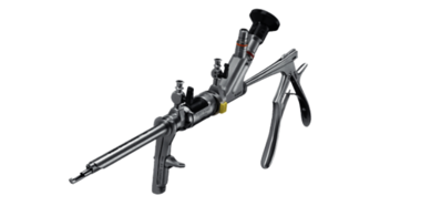
VERTEBRIS
stenosis
Two specialized instrument sets, designed for anterior and posterior approaches.
More information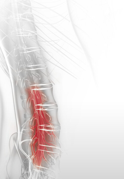

VERTEBRIS
lumbar
Designed for full-endoscopic decompression of the lumbar and thoracic spine.
More information
